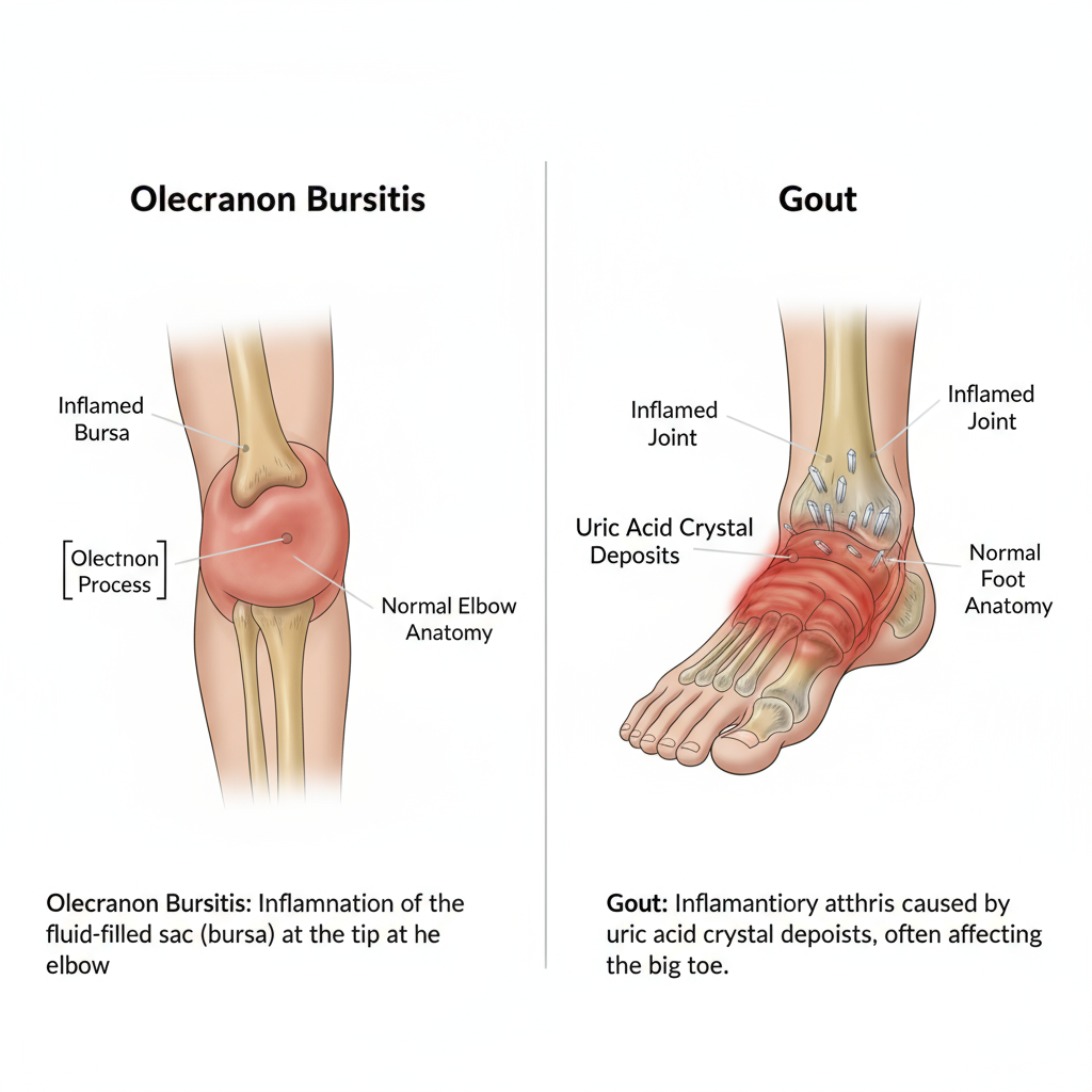
Olecranon bursitis and gout can both cause pain and swelling in the elbow, which can be confusing to diagnose. However, the two conditions have distinct causes, symptoms, and diagnostic methods.

Key Diagnostic Differences
- Causes
- Olecranon bursitis: Inflammation of the olecranon bursa, located at the tip of the elbow. It is usually caused by repetitive friction (e.g., leaning on a desk), direct trauma, or infection. It can also be secondary to systemic inflammatory conditions such as rheumatoid arthritis or gout.
- Gout: This condition occurs when high levels of uric acid in the blood cause uric acid crystals to accumulate around the joints, causing inflammation. Gout most commonly affects the big toe joint, but can also affect other joints, including the elbow.
- Symptoms and Characteristics
- Olecranon bursitis:
o Swelling: Swelling is characterized by a round, “golf ball” or “ping pong ball”-sized swelling at the tip of the elbow. This swelling is caused by fluid buildup within the bursa. o Pain: Pain increases when pressing on the swollen area or bending the elbow.
o Limited movement: Severe swelling may limit elbow movement.
o Warmth/redness: In infectious bursitis, the swollen area becomes red and hot, and may be accompanied by systemic symptoms such as fever and chills. - Gout:
o Pain attacks: Severe, sudden pain often begins at night. The pain can be so severe that the expression “even the breeze hurts” is used.
o Swelling/redness: The entire joint becomes swollen, red, and warm.
o Site: Gout usually affects one joint first, most commonly the big toe joint. The elbow can also develop, but this is not the typical gout site.
o Tophi: In chronic gout, “tophi,” nodules formed by uric acid crystals, may form around the elbow. 3. Diagnosis - Clinical Diagnosis:
o A physician makes the initial diagnosis based on a patient’s medical history (trauma, lifestyle habits, etc.) and a physical examination (location and type of swelling, location of pain, etc.).
o Olecranon bursitis is characterized by swelling limited to the elbow, whereas gout manifests inflammation throughout the joint. - Imaging Tests:
o X-ray: It is difficult to make a definitive diagnosis with X-ray alone for either condition. However, X-rays can help differentiate from similar conditions, such as fractures or other arthritis. Chronic gout may reveal joint deformities or tophi.
o Ultrasound/MRI: Useful for assessing inflammation or fluid retention in the bursa. - Most Definitive Diagnostic Method (Aspiration and Synovial Fluid Analysis):
o The most accurate differential diagnosis is to obtain fluid directly from the swollen area (aspiration) and analyze it.
o Olecranon bursitis: White blood cell count, Gram stain, and culture are performed to confirm infection.
o Gout: Uric acid crystals are identified under a microscope. Uric acid crystals are needle-shaped, polarized birefringent crystals and are crucial for the diagnosis of gout. - Blood Tests:
o Gout: Measuring blood uric acid levels is an important reference for diagnosing gout.
o Olecranon Bursitis: Infectious bursitis may present with elevated inflammatory markers (ESR, CRP) and white blood cell count.